Microscale Immune and Cell Analysis (MICA)
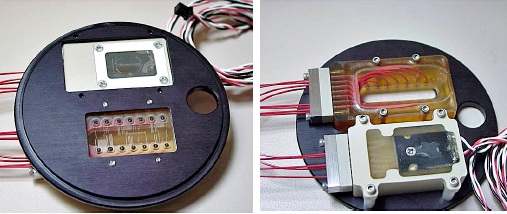
Researchers at Sandia National Laboratories have created a sophisticated, integrated platform for single-cell manipulation and interrogation. Called Microscale Immune and Cell Analysis (MICA), this platform represents a revolutionary breakthrough in biological research. MICA offers experimenters the ability to understand cell behavior at the molecular and cellular levels with unprecedented speed, resolution, sensitivity, and multiplexing.
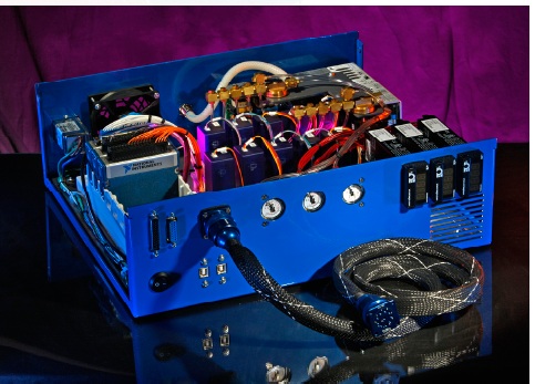
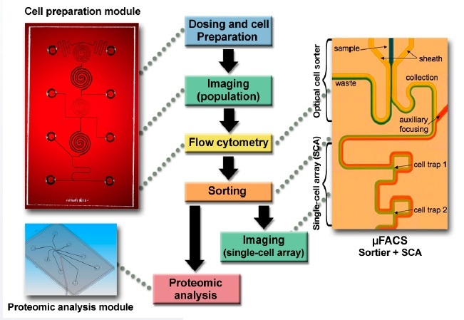
Unlike other lab-on-a-chip platforms, MICA helps researchers target, sort, and measure samples as small as a single cell in a precise, automated system that incorporates flow cytometry. And because MICA is based on microfluidics, it enables biological measurements that are impossible at a conventional scale. The promise of MICA is immense. MICA has already proven its value in a major Sandia research program that is investigating the responses of immune cells to pathogens. When fully developed as a research tool, the MICA platform may enable scientists to integrate experimentation on myriad cell types, thereby providing a systems understanding of multiple cellular mechanisms that have long eluded researchers. MICA helps researchers unlock the secrets of cell functions by offering an integrated platform of tools that include dosing, cell preparation, imaging, flow cytometry, sorting, and single-cell arrays (SCAs). These tools can be used to perform comprehensive biology experiments and measurements on samples as small as a single cell. MICA also offers high-sensitivity and high-throughput analyses that are both rapid and convenient. By using MICA, researchers can automate and integrate cellular experiments, thereby yielding rich—and sometimes previously unknown—findings.
MICA offers an array of benefits over conventional approaches:
- New functionality. MICA offers the capability to perform experimentation and measurement at single-cell resolution; this is not possible with commercial fluorescence-activated cell sorter (FACS) or proteomic techniques.
- Systems understanding. MICA is a single, unified platform that can perform multiple measurements (e.g., translocation and protein abundance measurements) on the same population of cells, enabling understanding of the entire system of cell responses.
- Speed and efficiency. Researchers can use MICA to measure multiple components of cellular pathways simultaneously and quickly—without requiring large quantities of cells and expensive reagents.
- Enhanced control. MICA’s systems integration capability eliminates manual steps and provides scientists with complete control of the cell environment—enabling measurements of concentration and time events.
- Convenient. MICA neatly fits on a typical microscope stage.
- Versatility. MICA can handle many different cell types, and the MICA hardware adapts to many commercial microscopes.
Within Sandia’s research of immune response, MICA has increased our understanding of exactly how immune cells counter viral or bacterial invasion. When fully developed, MICA could revolutionize the study of multiple cell mechanisms, engendering knowledge to advance biology, medicine, diagnostics, and therapeutics. Potential applications of MICA include the following:
- Drug discovery and therapeutics. High information-content screening, evaluating how drug candid
- Diagnostics. Observing and sorting abnormal cells, enriching rare cell types
- Personalized medicine. Monitoring the behavior of individual patient cells
- Immune and infectious disease. Observing the mechanisms of infection
- Biomarker discovery. Identifying and quantifying specific proteins (e.g., cytokine profiling)
- Srivastava N, Brennan JS, Renzi RF, Wu MY, Branda SS, Singh AK, Herr AE. Fully Integrated Microfluidic Platform Enabling Automated Phosphoprofiling of Macrophage Response. Analytical Chemistry 81, 3261-3269 (2009).
- Boas, G. (2008). Sorting it all out: Optical forces enable cell sorting with infectious agents. Biophotonics Research [Online]. August 2008.
- James, CD, N Reuel, ES Lee, RV Davalos, SS Mani, A Carroll-Portillo, R Rebeil, A Martino, C Apblett. (2008). Impedimetric and optical interrogation of single cells in a microfluidic device for real-time viability and chemical response assessment. Biosensors and Bioelectronics 23:845–861.
- Patel, KD, TD Perroud. (in preparation). The microflow cytometer. Optical Sorters. Pan Stanford Publishing: Hackensack, NJ. To be published in May 2009.
- Perroud, TD, JN Kaiser, JC Sy, TW Lane, CS Branda, AK Singh, KD Patel. (2008). Microfluidic-based cell sorting of Francisella tularensis infected macrophages using optical forces. Analytical Chemistry 80:6365–6372.
- Perroud, TD; Meagher RJ; Kanouff, MP; Renzi, RF; Wu, M; Singh, AK; Patel, KD. Isotropically Etched Radial Micropore for Cell Concentration, Immobilization, and Picodroplet Generation. Lab on a Chip 9, 507-515 (2009). Featured on journal back cover.
- Sinclair, MB, DM Haaland, JA Timlin, HDT Jones. (2006). Hyperspectral confocal microscope. Applied Optics 45:6283–6291.
SD# 11030.1; SD# 10838.1; SD# 10740.2, SD# 10978.2, SD# 11149; SD# 11152; SD# 8604.1; SD# 11162.1: SD# 10745.1; SD# 10545.1
Published11/16/2011
Last Updated1/7/2025