Large Field of View, 3D X-ray Phase Contrast Imaging System
A high-sensitivity X-ray Phase Contrast Imaging system for non-destructive 2D radiography and 3D tomography of low-density composite materials
Sandia National Laboratories has developed a high-sensitivity X-ray Phase Contrast Imaging (XPCI) system that provides a method for non-destructive 2D digital radiography and 3D tomography of low-density composite materials. Non-destructive evaluation and inspection is essential to establishing confidence in the functionality and security of a variety of components. Many of these parts contain low-density encapsulants to protect components from shock, thermal fluctuations, and high voltage breakdown. Sandia’s XPCI system incorporates unparalleled grating technology, which enables identification of defects in low-density materials that are usually transparent or indistinct using traditional X-ray imaging.
Sandia uses an innovative semiconductor manufacturing process that combines Bosch deep reactive ion etching (DRIE), atomic layer deposition (ALD), and pulse gold electroplating to create higher aspect ratio gratings with better uniformity and larger surface area— increasing field of view and sensitivity over the current state-of-the-art. This novel grating technology enabled the creation of a compact, bench-scale XPCI device with the ability to detect cracks, voids, or delamination in low density materials. Sandia’s XPCI system enables non-destructive inspection throughout the assembly process—improving overall confidence in part reliability. Sandia has also demonstrated advanced image processing techniques that enable extraction of features in low density materials in proximity to high density components such as foam microstructure in the presence of high-Z structures (e.g., wires). This novel XPCI system will increase confidence in components used in high consequence national security systems and has potential impact on medical imaging, homeland security, building and material safety, transportation safety, and food safety inspections.
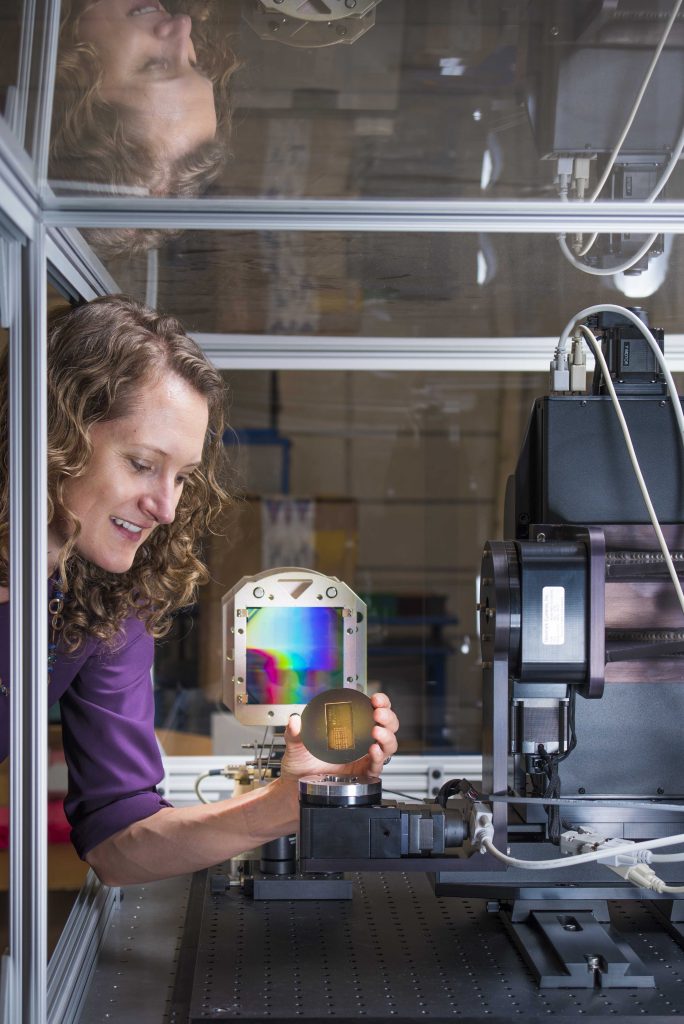
Image caption: Sandia National Laboratories researcher Amber Dagel holds a calibration sample to be loaded into the labs’ X-ray phase contrast imaging machine. Dagel is principal investigator for the labs’ work into using X-ray phase contrast imaging to study low-density materials.
- Large field of view
- Capable of 2D digital radiography and 3D tomography
- Benchtop system
- Improved sensitivity and defect detection in low density materials
- Reduced costs by identifying defects early
- Aging and state characterization
- Medical imaging
- Food safety inspections
- Lighting up the study of low-density materials
Sandia National Laboratories media release (July 17, 2017) - R&D 100 Market Disruptor Product Special Recognition: 2016
SD#12227
Published4/13/2018
Last Updated1/10/2019